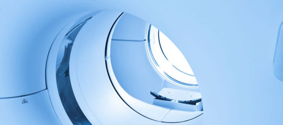
MRI
Magnetic resonance imaging (MRI) is a medical imaging technique used in radiology to form pictures of the anatomy and the physiological processes of the body. MRI scanners use strong magnetic fields, magnetic field gradients, and radio waves to generate images of the organs in the body. MRI does not involve X-rays or the use of ionizing radiation, which distinguishes it from CT or CAT scans.
MRA
MRA (Magnetic Resonance Angiography) is a specialized type of MRI exam used to obtain images of the arteries of the brain, carotid arteries, aorta, renal arteries and vessels of the extremities. MRA is used to detect problems with blood vessels that may be causing reduced blood flow.
MRI Arthrogram
An MRI arthrogram is an imaging study conducted to diagnose an issue within a joint. The exam is done in two parts and usually with the aid of a contrast agent called gadolinium that will help to highlight the visualization of joint structures and improve the MRI evaluation.
Arthrogram
- An arthrogram is used to:
- Identify the presence of abnormal growths or cysts.
- For diagnosis of complete rotator cuff tears, adhesive capsulitis, tear of the rotator interval, disorders of the biceps tendon and impingement syndrome.
CT
Computed Tomography (CT), also called a CAT scan, and is a medical imaging exam that takes computer assisted x-rays that produces two and three dimensional detailed images of the soft tissues and bones within the body.
CTA
Computed tomography angiography (also called CT angiography or CTA) is a computed tomography technique used to visualize arterial and venous vessels throughout the body. Using contrast injected into the blood vessels, images are created to look for blockages, aneurysms (dilations of walls), dissections (tearing of walls), and stenosis (narrowing of vessel).
CTU
CT urography (CTU) is used as primary imaging techniques to evaluate patients with blood in the urine (hematuria), follow patients with prior history of cancers of the urinary collecting system and to identify abnormalities in patients with recurrent urinary tract infections.
Nuclear Medicine
Nuclear Medicine imaging uses small amounts of radioactive material to diagnose and detect a full spectrum of diseases. The material is typically injected or swallowed and images are then acquired to aid in the diagnosis of your condition.
Nuclear SPECT
Single-photon emission computed tomography (SPECT, or less commonly, SPET) is a nuclear medicine tomographic imaging technique using gamma rays. It is very similar to conventional nuclear medicine planar imaging using a gamma camera (that is, scintigraphy). but is able to provide true 3D information. This information is typically presented as cross-sectional slices through the patient, but can be freely reformatted or manipulated as required.
Ultrasound
Ultrasound (also called a sonogram) is a medical imaging exam that uses sound waves to create images. These images show the structure and movement of the body’s organs and blood flow.
X-ray
An X-ray is a non-invasive exam that provides valuable information to help physicians diagnose and treat medical conditions. Imaging with x-rays involves using a small dose of radiation to produce images of the inside of the body. X-rays are the oldest and most frequently used form of medical imaging.

